


Senior Investigator Research Interests
Branching morphogenesis is a fundamental attribute of many organ systems. The laboratory of Dr. Yosuke Mukoyama (Yoh-suke Mukouyama in PubMed) aims to uncover the molecular control of the morphologic processes underlying the patterning of tubular branching networks, particularly the vascular and nervous systems, which share several anatomic and functional characteristics and are often patterned similarly in peripheral tissues. These characteristics suggest that these two networks are interdependent during tissue development and homeostasis. Thus, Dr. Mukoyama is studying neuronal influences on vascular branching patterns and vascular influences on both neuronal guidance and neural stem cell maintenance. His laboratory approaches these problems using a combination of high-resolution whole-mount imaging, molecular manipulations, advanced genetic perturbations, next-generation sequencing, and in vitro organ culture techniques.
By developing a model system of skin vasculature that allows for the direct observation of congruent vascular and neural networks in an anatomically recognizable pattern, Dr. Mukoyama and his colleagues have begun to unravel the neural control of vascular development. The CXCL12–CXCR4 and VEGF-A–VEGFR2/Nrp1 signaling pathways are important for sensory nerve-mediated arterial branching, while NGF–TrkA signaling is essential for guiding sympathetic axons along blood vessels. Using high-resolution whole-mount imaging in vivo Ca2+ indicator mice, they are currently examining how sensory activity changes in mutants with neurovascular mispatterning in developmental and pathological contexts, including obesity-related nerve disorders. These studies will provide novel conceptual advances in the development and maintenance of functional neurovascular wiring.
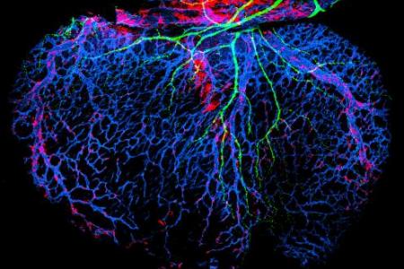
Just as nerves influence vascular patterns in the skin, Dr. Mukoyama and his colleagues have also shown that blood vessels influence nerve patterns in the developing heart. Using whole-mount imaging, they demonstrated that sympathetic axons branch alongside large-diameter coronary veins in the sub-epicardial layer of the dorsal ventricular wall before innervating final targets such as coronary arteries and cardiomyocytes in the deeper myocardial layer. This novel neurovascular association is established by sequential expression of NGF in the subepicardial venous vascular smooth muscle cells (VSMCs) and myocardial arterial VSMCs of coronary vessels. Using ultra-high-frequency non-invasive imaging, they found abnormal cardiac functions, such as irregular cardiac rhythms, in mutant embryos lacking the autonomic nervous system. Using 2D co-culture, 3D organoids and assembloids, and advanced imaging, they are currently examining what happens to myocardial development and conduction system architecture in the absence of the autonomic nervous system and how autonomic nerves affect cardiomyocyte differentiation and maturation. These studies will provide a mechanistic framework for the functional consequences of sympathetic innervation in the developing heart.
No examination of neurovascular interactions could neglect the brain. The vasculature component of this specialized niche microenvironment helps to retain neural stem cell potential, while the CNS parenchyma develops a blood-brain barrier (BBB) and lacks a lymphatic system. Dr. Mukoyama and his colleagues have also found BBB disruption and abnormal expression of the lymphatic endothelial transcription factor Prox1 in tumor-associated endothelial cells in glioma and metastatic tumors. Ongoing work investigates the relationship between ectopic Prox1 expression and BBB dysfunction.
These three independent but thematically interrelated projects provide unique opportunities to advance understanding of the dynamic features of both vascular and neuronal branching networks.
Images

Sympathetic innervation of dermal blood vessels
Whole-mount double immunofluorescence confocal microscopy with antibodies to the sympathetic axon marker tyrosine hydroxylase (TH, green) and the vascular smooth muscle cell (VSMC) marker αSMA (red) shows the sympathetic innervation of VSMC-covered blood vessels in the dermis of the dermis of mouse limb skin at embryonic (E) day 18.5 (Li et al. Biol Open 13: bio060147).

A unique expression of hepatic leukemia factor (Hlf) in the cephalic paraxial mesenchyme
Triple RNA whole-mount in situ hybridization chain reaction of E8.5 wild-type embryos revealed the expression of Hlf (green), as well as the paraxial mesoderm markers Pax3 (blue) and Eomes (red) in the cephalic region. Koui et al. showed that Hlf-expressing cephalic paraxial mesenchymal cells contribute to the developing brain vascular cells such as endothelial cells and pericytes (Koui et al. Biol Open 11: bio059510, 2022).

Lymphatic vessels in the postnatal brain meninges.
Using the Prox1-GFP BAC transgenic reporter (green) and immunostaining with antibodies to lymphatic markers LYVE-1 (red) and pan-blood vessel marker PECAM-1 (blue), we have carried out whole-mount imaging of dural lymphatic vasculature at postnatal stages. We have found that between birth – postnatal day 13 (P)13, lymphatic vessels extend alongside dural blood vessels from the side of the skull towards the midline. The image was published in Developmental Dynamics (Izen et al. Dev Dyn 247:741-753, 2018).

Structural interdependence of blood vessels and nerves to form coordinated branching networks
EC, endothelial cells; VSMC, vascular smooth muscle cells. Modified from Fig. 3 in James & Mukouyama. Semin Cell Dev Biol 22: 1019-1027, 2011.

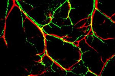
Nerve-artery alignment in developing skin
Sensory nerve (green) align with arteries (red) in the developing mouse skin. The image was published in Cell (Mukouyama et al. : 693-705, 2002).

Chemokine controls nerve-vessel co-patterning
Chemokine receptor expression (red) was restricted to nerve (green)-associated + arteries (blue). The image was published in Developmental Cell (Li et al. Dev Cell 24: 359-371, 2013).

Cardiac axons follow coronary vessels in embryonic heart
Cardiac sympathetic axons (green) follow VSMC-associated large-diameter coronary vessels in the subepicardium of the dorsal ventricular wall of the developing mouse heart. The image was selected as the cover of the issue in Development (Nam et al. Development 140: 1475-1485, 2013).

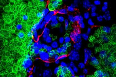
Intra-neural hemorrhage in the brain: a mouse model of human CCMs
The vascular lumen appears to have ruptured resulting in hemorrhage in conditional mutants as evidenced by erythrocytes (green) outside the vessel enclosure (red). The image was published in Human Molecular Genetics (Cunningham et al. Hum Mol Genet 20: 3198-3206, 2011)

Venous expression of EphB1 in embryonic limb skin vasculature
Whole-mount triple immunofluorescence analysis of forelimb skin of .5 embryos was performed with antibodies to (green) in combination with the pan-endothelial cell marker PECAM-1 (blue) and the vascular smooth muscle cell (VSMC) marker alpha SMA (red). By .5, arterial vessels are densely covered with VSMCs, whereas less or no VSMC coverage occurs in venous vessels. expression is restricted to the venous vasculature. No expression is detectable in the arterial and lymphatic vasculature. The image was published in Developmental Dynamics (Li et al. Dev Dyn 2013).

Skin lymphangiogenesis
Whole-mount confocal microscopy reveals that + (blue)/LYVE-1+ (green) lymphatic vessels sprout toward the dorsal midline in .5 mouse dorsal skin. PECAM-1+ blood vessels (red) have reached the midline at this stage. The image was published in Development (James et al. Development 2013).

Gut angiogenesis and neurogenesis
An embryonic day (E)15.5 whole-mount small intestine stained with antibodies to PECAM1 (blood vessels, blue), TuJ1 (neuronal tubulin, neurons, green), and tyrosine hydroxylase (sympathetic neurons, red). Here, the enteric nervous system (ENS, green) can be seen surrounding the full length of the small intestine. Sympathetic nerves (red and green) project along arteries in the mesentery until they reach the intestinal wall. Upon reaching the ENS, these nerves defasciculate and extend throughout the existing enteric nerve plexus. The image was published in Developmental Dynamics (Hatch & Mukouyama, Dev Dyn 2014).

Sema3s-Nrp2 signaling is required for skin lymphangiogenesis
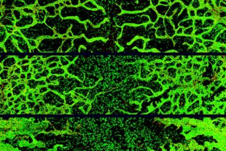
High-resolution whole-mount staining of back skin from E15.5 mutants and control littermate was performed with antibodies to lymphatic markers PROX1 (red) and LYVE1 (green). Sema3f;Sema3g double mutants exhibit increased lymphatic branching (middle image) compared with control littermate (top image). Nrp2 mutants exhibit decreased lymphatic branching and increased lymphatic endothelial cell growth. The image will be published in Biology Open (Uchida et al. Biol Open 2015).

Adult ear skin innervation and vascularization
High resolution whole-mount adult ear skin stained with antibodies to PECAM1 (blood vessels, blue), TuJ1 (neuronal tubulin, neurons, green), and alpha SMA (vascular smooth muscle cells, red). The alignment of Tuj1+ peripheral nerve bundles with large- or middle-diameter blood vessels covered with alpha SMA+ vascular smooth muscle cells. The image was the 8th place in the 2015 Nikon Photomicrography competition.

SV patient with Glenn physiology and APCs who had increased angiogenic activity in vitro
The image will be published in the Journal of Thoracic and Cardiovascular Surgery (Sandeep et al. J Thorac Cardiovasc Surg. 2015).

Some pericytes are of hematopoietic origin in embryonic skin vasculature
We discovered that the developmental sources of dermal pericytes are heterogeneous and a portion of dermal pericytes has a hematopoietic/myeloid origin. Confocal image obtained from fate-mapping experiments using embryo that have pan-hematopoietic Vav-Cre and R26R-EYFP reporter demonstrates that some NG2+ pericytes (red) associated with PECAM-1+ blood vessels (blue) are also EYFP+ (green), sugesting that EYFP+NG2+ pericytes originate from hematopoietic cells. The image was published in Cell Reports (Yamazaki et al. Cell Rep 2017).

Soluble APP negatively regulates NSC growth in culture
Whole-mount immunostaining of SVZ-derived neurospheres with antibodies to a NSC marker Sox2 (red) and a proliferation marker phospho-histone H3 (green). The treatment of soluble amyloid precursor protein (right) suppresses NSC growth in culture, compared to that of control alkaline phosphatase protein (left). The image was published in Development (Sato et al. Development 144: 2730-2736, 2017).

High resolution whole-mount tissue-cleared mouse embryo
High resolution whole-mount tissue-cleared mouse embryo (embryonic day 13.5) stained with antibodies to PECAM1 (blood vessels, blue) and TuJ1 (neuronal tubulin, neurons, green). The image was selected in the top 100 in the 2019 Nikon Small World competition.

High resolution whole-mount tissue-cleared mouse gut
High resolution whole-mount tissue-cleared mouse gut (embryonic day 13.5) stained with antibodies to PECAM1 (blood vessels, blue), TuJ1 (neuronal tubulin, neurons, red) and TH (tyrosine hydroxylase, sympathetic neurons, green). The image was selected in the top 100 in the 2019 Nikon Small World competition.

Whole-mount and ultrasound analysis of mutants lacking autonomic nerves
Whole-mount double immunofluorescence confocal microscopy was performed with the pan-neuronal marker Tuj1 (class III b-tubulin, green) and endothelial cell marker PECAM-1 (red). The Phox2b-/- ventricle has a near complete absence of autonomic neurons. M-mode ultrasound analysis of a Phox2b-/- embryo shows irregular ventricular contraction (arrows) both in rhythm and magnitude of contraction suggesting ectopic atrial or ventricular focus. The images were published in Ultrasound in Medicine & Biology (Mokshagundam et al. Ultrasound Med Biol 47:751-758, 2020).

Coronary vasculature in the heart from Cxcr4-overexpression embryos
Whole-mount immunofluorescence confocal microscopy of E15.5 heart was performed with antibodies to pan-endothelial cell marker PECAM-1 (green) along with venous and capillary marker EMCN (red) to visualize coronary veins in the dorsal subepicardial layer of the ventricular wall. Endothelial cell-specific Cxcr4 overexpression embryos fail to form hierarchical coronary vascular network. The image was selected as the cover of the issue in Developmental Biology (Li et al. Dev Biol 477: 70-84. 2021).

Sympathetic neurons contribute to iPSC-derived cardiomyocyte maturation
Double immunofluorescence confocal microscopy demonstrated the co-culture of EGFP-expressing iPSC-derived cardiomyocytes (white) with tyrosine hydroxylase-expressing sympathetic neurons (green) isolated from mouse embryos (Kowalski et al. Front Cell Dev Biol 10: 850645. 2022).

Mouse ES-derived vascular organoids, transplanted onto chick CAM, exhibit a hierarchical vascular network
PECAM-1+ vessels (blue) displayed a branched hierarchy with a greater number of associated NG2+ pericytes (red). Large-diameter vessels wrapped with αSMA+ vascular smooth muscle cells (green) developed within the organoid (arrowheads) (Kowalski et al. Sci Rep 15: 7150. 2025).

Diet-induced obesity (DIO) enhances capsaicin-mediated acute pain
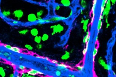
Nociceptive pain stimuli evoke forelimb wiping responses (left) and sensory activation (right) in DIO mice. Local interaction between keratinocytes (magenta) and nociceptive sensory nerves (green) in DIO epidermis (upper right) via NGF-TrkA-PI3K signaling axis promotes the initiation and progression of painful small fiber neuropathy. Further interaction between skin nociceptive sensory nerves (green) and blood vessels (blue, capillary; blue + red, artery and vein) may facilitate sensory hypersensitivity in the DIO skin (middle and lower right) (Koui et al. iScience 28: 112047. 2025).

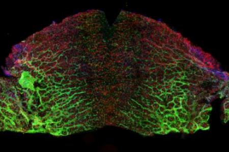
Endothelial PROX1 induces blood-brain barrier disruption in the central nervous system
Ectopic PROX1 expression in brain endothelial cells disrupts the blood-brain barrier (BBB), leading to vascular leakage by inhibiting key BBB-associated gene expression and Wnt/β-catenin signaling. The cover image highlights the BBB permeability phenotype in a mouse with PROX1 overexpression, showing leakage of dextran (red) and endomucin-labeled capillaries (cyan). The image was selected as the cover of the issue in JCI insight (González-Hernández et al. JCI insight 11: e187716. 2026).

Defective co-patterning of sensory nerves and blood vessels in Vhl mutants
Whole-mount immunofluorescence confocal microscopy was performed on E15.5 limb skin from endothelial cell-specific von Hippel-Lindau (Vhl) mutants (right panel) and their control littermates (left panel). Samples stained for PECAM-1 (magenta, endothelial cells) and Tuj1 (green, axons) revealed defective co-patterning of sensory nerves and blood vessels in Vhl mutants. The image was selected as the cover of the issue in Development (Li et al. Development 153: dev204519).









Meet the Team

Yosuke Mukoyama, Ph.D.
Yoh-suke Mukouyama, whose legal name is Yosuke Mukoyama, publishes as Yoh-suke Mukouyama. He received his A.B. in pharmacy studies from Tokyo University of Science and his M.S. and Ph.D. in developmental biology from the University of Tokyo. As a student in the laboratory of Dr. Atsushi Miyajima, he studied the properties of embryonic endothelial cells and hematopoietic stem cells (HSCs). He did postdoctoral research in the laboratory of Dr. David Anderson at the California Institute of Technology where he demonstrated that peripheral nerves direct arterial organization and formation by secreting VEGF; he also studied the properties of neural stem cells (NSCs). He joined the NHLBI as a tenure-track Investigator in 2006. He also serves on the editorial board of Developmental Dynamics and is a member of the North American Vascular Biology Organization.
Contact the lab

Krista Gill, B.S.

Wenling Li, Ph.D.

William Kowalski, Ph.D.

Sara González Hernández, Ph.D.

Sarah Ruttenberg, Ph.D.
Josephine Watkins, B.S.

Jonathan Shin, B.S.
Natalie Rowland, B.S.
