
MORE INFORMATION
Stents Getting a Stent
Having a stent placed is a minimally invasive procedure, meaning it does not require a large open incision.
Coronary and carotid artery stenting
Procedures to place a stent to treat coronary and are similar. In both, your healthcare provider will use catheterization to thread a thin tube with a stent and an empty balloon on the end through the blood vessel to the narrowed or blocked artery. Once the balloon is in place, filling it with air opens the stent to hold up the artery walls. Then your provider will empty the balloon of air again and remove the tube and the empty balloon from the artery.
- Percutaneous coronary intervention (PCI), or coronary , opens narrowed or blocked arteries of the heart to treat coronary heart disease. A coronary stent can be placed during a PCI to prevent the artery from narrowing again. Sometimes the procedure is done in an emergency, such as during a heart attack.

- Carotid artery stenting is a minimally invasive treatment for severe carotid artery disease. Usually, the procedure to place a carotid stent includes temporarily blocking the blood flow from the narrowed artery to the brain. This helps prevent any pieces of from reaching the brain and causing a stroke.

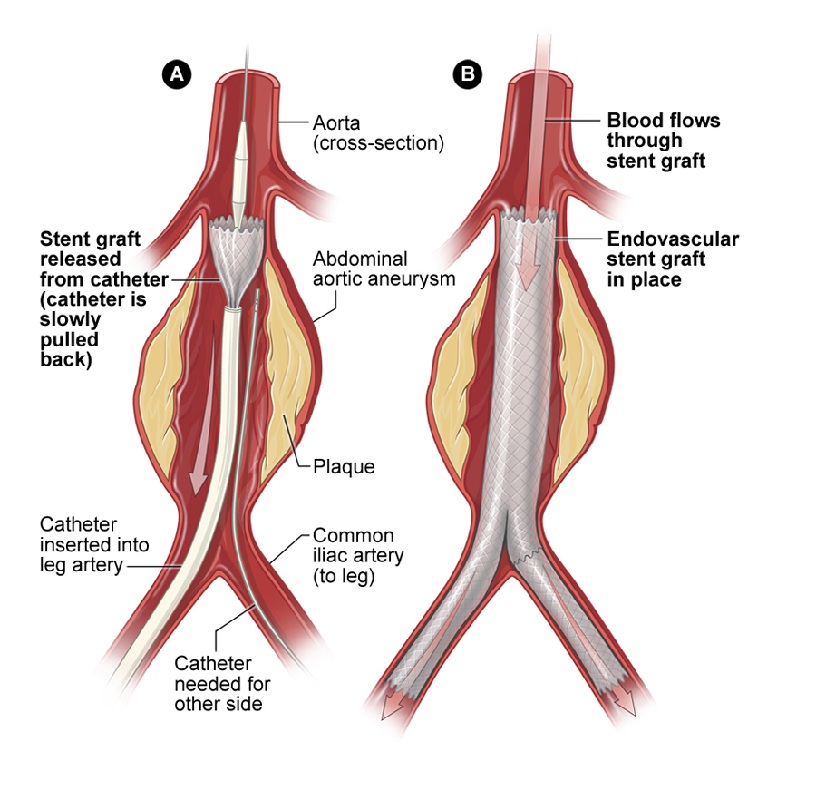
Aortic aneurysm stenting
The procedure to place a stent graft in the aorta to treat an aneurysm is called endovascular aneurysm repair. Your healthcare provider inserts a stent graft through a large blood vessel using a catheter and guides it up your arteries to the location of the aneurysm. Once it is in the right place, the stent graft opens up and attaches to the walls of the aorta. This will seal off the aneurysm and prevent it from getting bigger or rupturing. After placing the stent graft, your provider may inject a contrast dye into your blood and take an X-ray of your aorta to make sure blood is not leaking into the aneurysm.
Airway stenting
Bronchoscopy uses a small camera on the end of a long tube to look inside the airways in your lungs. To do that, your healthcare provider slides the camera, or bronchoscope, through your nose or mouth and down your throat into the airways. During the procedure, your provider can also insert an airway stent or take samples for diagnostic tests.
If you need a stent, your provider will slide a thin tube that carries the folded stent along the side of the bronchoscope. Your provider may also use fluoroscopy, a type of X-ray imaging, or ultrasound to help guide stent placement. Once it is in the right area, the stent will expand in the narrowed airway to push it open. After placing the stent, your provider may check your lungs using a chest X-ray.