Congenital Heart Defects
0
Congenital Heart Defects

Congenital Heart Defects Types

There are many types of congenital heart defects. They range from simple to complex and critical.

  • Simple defects may get better on their own without surgery. Sometimes, a baby with a simple defect will not have any symptoms . Examples of simple heart defects include atrial and ventricular septal defects, patent ductus arteriosus, and pulmonary stenosis .
  • Complex and critical defects may cause life-threatening symptoms that require immediate treatment. An example of a critical heart condition is the Tetralogy of Fallot. Babies born with this or another critical congenital heart defect typically have low levels of oxygen soon after birth and need surgery within the first year of life.

Atrial septal defect

An atrial septal defect is a hole in the wall of the heart between the left and right atria, which are the two upper chambers of the heart. The hole causes blood to flow from the left atrium and mix with the right atrium, instead of going to the rest of the body. An atrial septal defect is considerered a simple congenital heart defect because the hole may close on its own as the heart grows during childhood.

Cross-section of a normal heart and a heart with an atrial septal defect.
Cross-section of a normal heart and a heart with an atrial septal defect. Figure A shows the structure and blood flow inside a normal heart. The blue arrow shows the flow of oxygen-poor blood as it is pumped from the body into the right atrium and then to the right ventricle. From there, it pumps through the pulmonary artery to the lungs, where it picks up oxygen. The oxygen-rich blood, shown by the red arrows, flows from the lungs through the pulmonary veins into the left atrium. Figure B shows a heart with an atrial septal defect. The hole allows oxygen-rich blood from the left atrium to mix with the oxygen-poor blood from the right atrium. The mixed blood is shown with a purple arrow.

Ventricular septal defect

A ventricular septal defect is a hole in the wall between the left and right ventricles, which are the two lower chambers of the heart. Blood may flow from the left ventricle and mix with blood in the right ventricle, instead of going to the rest of the body. If the hole is large, the heart and lungs may need to work harder to pump blood. In addition, it may cause fluid to build up in the lungs.

Cross-section of a normal heart and a heart with a ventricular septal defect.
Cross-section of a normal heart and a heart with a ventricular septal defect. Figure A shows the structure and blood flow inside a normal heart. The blue arrow shows the flow of oxygen-poor blood as it is pumped from the body into the right atrium and then to the right ventricle. From there, it pumps through the pulmonary artery to the lungs, where it picks up oxygen. The oxygen-rich blood, shown with a red arrow, flows from the lungs through the pulmonary veins into the left atrium. Figure B shows two common locations for a ventricular septal defect. The defect, or hole, allows oxygen-rich blood from the left ventricle to mix with oxygen-poor blood in the right ventricle before the blood flows into the pulmonary artery. The mixed blood is shown with a purple arrow.

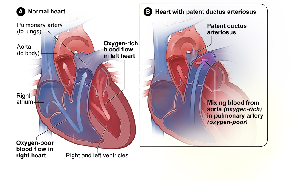
Patent ductus arteriosus

This common type of simple congenital heart defect occurs when a connection between the heart’s two major arteries, the aorta and the pulmonary artery, does not close properly after birth. This leaves an opening through which blood can flow when it should not. In many cases, small openings may close on their own.

Normal heart and heart with patent ductus arteriosus.
Normal heart and heart with patent ductus arteriosus. Figure A shows the interior of a normal heart and normal blood flow. The blue arrow shows the flow of oxygen-poor blood as it is pumped from the body into the right atrium and then to the right ventricle. From there, it pumps through the pulmonary artery to the lungs, where it picks up oxygen. The oxygen-rich blood, shown with a red arrow, flows from the lungs through the pulmonary veins into the left atrium. Figure B shows a heart with patent ductus arteriosus. The defect connects the aorta with the pulmonary artery, a connection that should have closed to form the ligamentum arteriosum (see Figure A) at birth. The hole allows oxygen-rich blood from the left atrium to mix with the oxygen-poor blood from the right atrium. The mixed blood is shown with a purple arrow.

Pulmonary stenosis

Pulmonary stenosis is a type of heart valve disease in which the pulmonary valve is too narrow or stiff. This affects how well blood can move from the heart to the pulmonary artery, the blood vessel that connects the heart to the lungs. Many children with pulmonary stenosis do not need treatment.

Tetralogy of Fallot

Tetralogy of Fallot is the most common complex congenital heart disorder and is a combination of four defects:

  • Pulmonary stenosis
  • A large ventricular septal defect
  • An overriding aorta. With this defect, the major blood vessel that carries blood to the body (aorta) is out of place. Instead of being above the left ventricle , it is located between the two ventricles. As a result, oxygen -poor blood from the right ventricle can flow directly into the aorta instead of into the blood vessel that carries blood to the lungs (pulmonary artery)
  • Right ventricular hypertrophy is when the heart has to work harder than normal which makes the muscle of the right ventricle thicker than normal
Cross-Section of a Normal Heart and a Heart with Tetralogy of Fallot.
Cross-Section of a Normal Heart and a Heart with Tetralogy of Fallot. Figure A shows the structure and blood flow inside a normal heart. The blue arrow shows the flow of oxygen-poor blood as it is pumped from the body into the right atrium and then to the right ventricle. From there, it pumps through the pulmonary artery to the lungs, where it picks up oxygen. The oxygen-rich blood, shown with a red arrow, flows from the lungs through the pulmonary veins into the left atrium. Figure B shows a heart with the four defects of tetralogy of Fallot, which includes pulmonary stenosis, ventricular septal defect, an overriding aorta, shown in the figure as increased output in the aorta; and a thickened right ventricular hypertrophy. These defects can cause the heart to work harder or allow oxygen-rich blood to mix with oxygen-poor blood. The mixed blood is shown with a purple arrow.

Other critical congenital heart defects

There are other types of critical congenital heart defects including:

  • Coarctation of the aorta
  • Double-outlet right ventricle
  • D-transposition of the great arteries
  • Ebstein’s anomaly
  • Hypoplastic left heart syndrome
  • Interrupted aortic arch
  • Pulmonary atresia with intact ventricular septum
  • Single ventricle
  • Total anomalous pulmonary venous return
  • Tricuspid atresia
  • Truncus arteriosus


Last updated on